


Komaba Campus
Graduate SchoolGraduate School of Engineering - Electrical Engineering and Information Systems
DepartmentDepartment of Electrical and Electronic Engineering
Photonics & Wireless Field
Measurement engineering
Photonics
Biosensing/imaging
Unveiling living systems with cutting-edge pulsed lasers
Ozeki Laboratory is pushing the boundaries of biological imaging using advanced optical pulse techniques. Our research focuses on developing new light sources and measurement systems, as well as applying these techniques to observe living systems. Our latest work includes high-speed molecular imaging, highly multiplexed imaging, and using quantum optics to increase sensitivity.
Research field 1
Research Overview
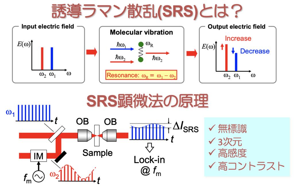
Ozeki Laboratory is using cutting-edge optical pulse and quantum technologies to visualize living systems and unlock the secrets of biological molecules. Our stimulated Raman scattering (SRS) microscopy technique (shown in the figure on the right) detects biological molecules based on molecular vibrational spectroscopic signatures, and we are constantly working to improve its performance and apply it to biological imaging. Our research covers a wide range of topics, from measurement principles to laser sources, control systems, optical systems, and data analysis methods. Our dedicated team of researchers is committed to exploring the boundaries of interdisciplinary research at the intersection of physics, chemistry, biology, and medicine, using our expertise in electronic engineering to push the limits of what is possible. Join us on our exciting journey of scientific exploration and innovation!
Research field 2
SRS spectrum microscope system
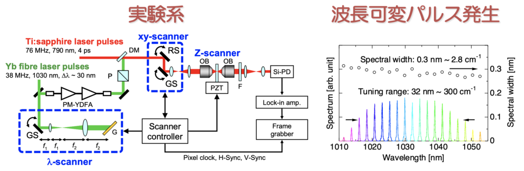
Unlocking the secrets of biological molecules requires the ability to visualize them with high precision and accuracy. In the past, conventional SRS microscopes had difficulty in distinguishing between multiple types of biological molecules. However, we have developed a solution: high-speed wavelength-switchable laser light sources that enable the acquisition of SRS images at various molecular vibration frequencies in just a few to tens of seconds. This breakthrough in SRS microscopy has made it possible to image multiple types of biological molecules, opening up new possibilities for exploring the intricate workings of living systems [1][2].
[1] Y. Ozeki et al., Opt. Lett. 37, 431 (2012).
[2] Y. Ozeki et al., Nature Photon. 6, 845 (2012).
Research field 3
Large-scale cell imaging
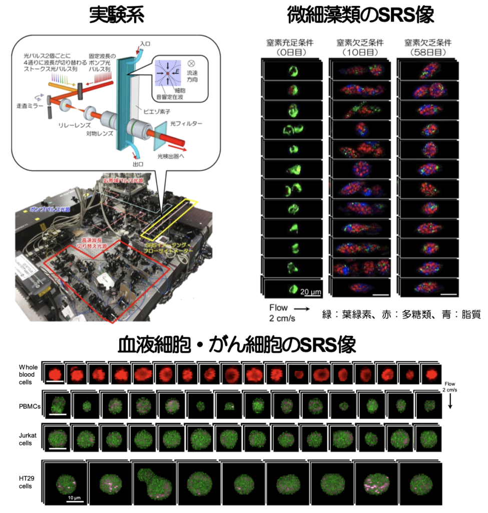
To realize SRS measurements of numerous cells, we have developed an ultrafast wavelength-switching light source and demonstrated multi-color SRS imaging of cells in high-speed flow [1]. With this technology, we can image biological molecules contained in each of the 10,000 or more cells, enabling imaging and measurement of nutrient distribution in microalgae and unlabeled measurements of blood cells and cancer cells. [1] Y. Suzuki et al., Proc. Natl. Acad. Sci. U.S.A. 116, 15842 (2019).
Research field 4
Pushing the sensitivity limit with quantum optics
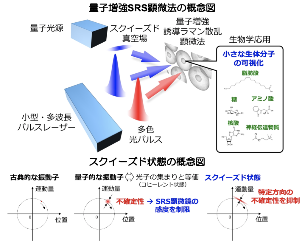
We are advancing the use of quantum optics to achieve ultra-high sensitivity in SRS microscopy. The signal-to-noise ratio in current SRS microscopy is limited by the quantum fluctuations (vacuum field fluctuations) of light. To reduce this fluctuation and improve the signal-to-noise ratio of SRS signals, we are introducing quantum-mechanical light known as squeezed light into SRS microscopy [1]. Our goal is to enhance the sensitivity of SRS microscopy and unlock new possibilities for exploring the intricate workings of biological systems. [1] Y. Ozeki et al., J. Opt. Soc. Am. B 37, 3288 (2020).